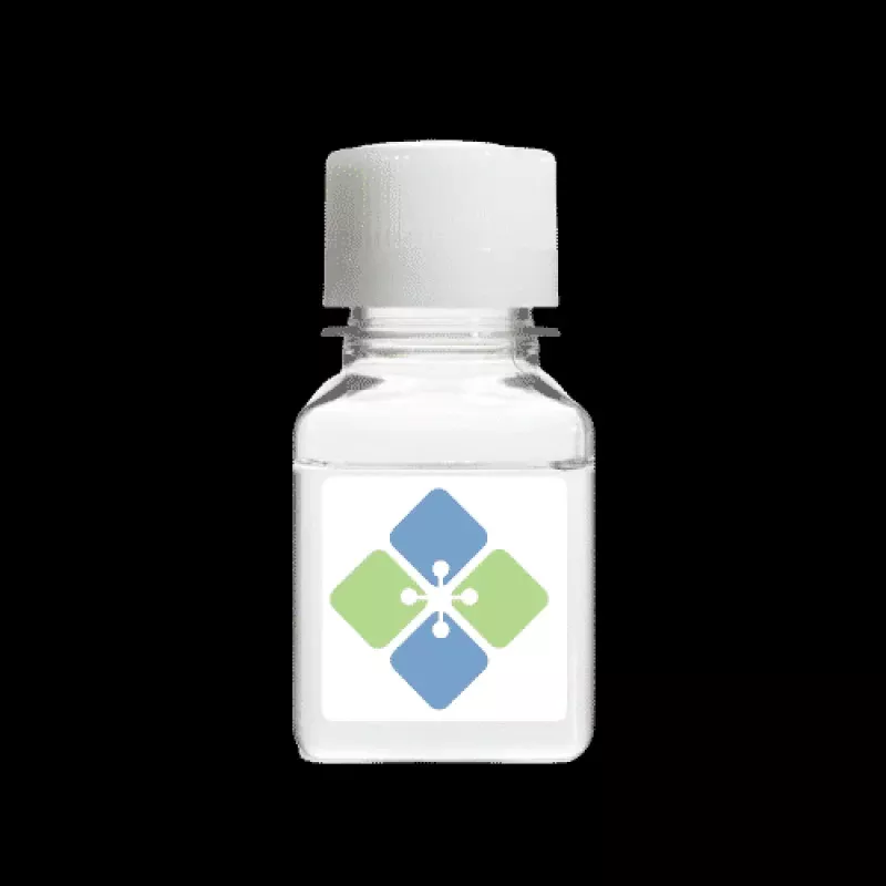
SDS-PAGE Destaining Solution Research Use Only
SDS-PAGE Destaining Solution is a biotechnology-grade methanol and acetic acid gel destaining reagent supplied as a 100 mL clear solution for research applications involving protein electrophoresis, Coomassie blue destaining, gel imaging, and analytical protein workflows. This ready-to-use solution is formulated to remove excess background stain from SDS-PAGE gels while preserving visible protein band contrast for downstream analysis. It is especially useful in workflows related to protein expression studies, electrophoretic separation, densitometry, western blot preparation, and routine protein visualization.
| Catalog number: | B2010020B |
| Lot number: | Batch Dependent |
| Expiration date: | Batch dependent |
| Volume: | 100 mL |
| Content Methanol and acetic acid solution | |
| Staining Time: | 1 hour to overnight |
| Appearance: | Clear Solution |
| Application: | SDS PAGE gels destaining |
| Shelf-Life: | Two years from date of manufacture |
| Storage: | Room temperature |
| Keywords: | Coomassie blue destaining buffer, gel destaining buffer, SDS-PAGE destaining reagent, coomassie blue destaining, coomassie destain, SDS Page destaining Solution |
| Grade: | Biotechnology grade. All components are highly pure (minimum 99%). All solutions are made with Type I ultrapure water (resistivity>18 M-cm) and are filtered on a 0.22 um. |
Scientific Overview
Destaining is a critical step in SDS-PAGE protein visualization workflows, particularly after Coomassie blue staining. Effective destaining removes excess background dye from the gel matrix while maintaining clear protein band visibility, helping researchers achieve stronger contrast and more interpretable electrophoresis results.
In laboratory settings, SDS-PAGE destaining solution is commonly used during protein purification analysis, expression verification, band visualization, densitometric comparison, and general gel documentation workflows. A properly balanced methanol/acetic acid solution helps reduce background staining and supports cleaner imaging for both routine bench work and more analytical protein characterization workflows.
Applications
- SDS-PAGE gel destaining after Coomassie blue staining
- Protein band visualization and contrast enhancement
- Protein expression and purification workflow analysis
- Gel documentation and densitometry preparation
- General electrophoresis and protein biochemistry workflows
Usage & Handling Guidance
Store at room temperature as specified. Use directly as a ready-to-use destaining reagent for stained SDS-PAGE gels. Destaining time may vary depending on gel thickness, stain intensity, and the desired background clarity. Researchers should monitor gels visually and adjust incubation time as needed for the intended imaging or analytical application.
- Preparation: Ready to use directly from bottle
- Handling: Use in a well-ventilated lab environment and according to standard chemical handling practices
- Optimization: Adjust destaining duration depending on gel background and desired contrast
- Workflow note: Gentle agitation may improve destaining consistency across the gel surface
What You Get
- 100 mL SDS-PAGE Destaining Solution
- Ready-to-use methanol/acetic acid gel destaining reagent
- Biotechnology-grade solution for protein gel workflows
- Suitable for Coomassie blue-stained SDS-PAGE gels
- For research use only (RUO)
Why Researchers Choose It
- Helps reduce gel background for cleaner protein band visibility
- Supports routine SDS-PAGE and protein analysis workflows
- Ready-to-use format simplifies bench preparation
- Useful for imaging, documentation, and densitometry workflows
- Room-temperature storage supports convenient lab handling
Frequently Asked Questions (FAQ)
- What is SDS-PAGE Destaining Solution used for?
It is used to remove excess Coomassie blue stain from SDS-PAGE gels so protein bands can be visualized more clearly. - Why is destaining important after Coomassie staining?
Destaining reduces background dye in the gel, which improves contrast and makes protein bands easier to analyze and document. - How long should gels be destained?
According to the supplied product information, gels may be destained for approximately 1 hour to overnight depending on the gel and desired background clarity. - Can this reagent be used before imaging or densitometry?
Yes. It is commonly used prior to gel imaging, documentation, and densitometric analysis to improve band visibility and reduce background interference. - Is this product intended for diagnostic or therapeutic use?
No. This product is supplied strictly for research use only and is not intended for diagnostic or therapeutic applications.
This product is for Research Use Only (RUO). It is not intended for diagnostic or therapeutic use.
References
- Fernandez-Patron C, Hardy E, Sosa A, Seoane J, Castellanos L. Double staining of coomassie blue-stained polyacrylamide gels by imidazole-sodium dodecyl sulfate-zinc reverse staining: sensitive detection of coomassie blue-undetected proteins. Anal Biochem. 1995 Jan 1;224(1):263-9.
- Brum AM, Thomas AD, Sabeur K, Ball BA. Evaluation of Coomassie blue staining of the acrosome of equine and canine spermatozoa. Am J Vet Res. 2006 Feb;67(2):358-62.
- Marshall T, Williams KM. Total protein determination in urine: elimination of a differential response between the coomassie blue and pyrogallol red protein dye-binding assays. Clin Chem. 2000 Mar;46(3):392-8.
- Lim CW, Chisnall WN, Stokes YM, Pratt R, Crooke MJ. Effects of sodium dodecylsulphate, dye concentration and paraprotein on coomassie blue dye-binding assays for protein in urine. Clin Biochem. 1988 Oct;21(5):277-81.
- Fujita T, Toda T, Ohashi M. Enzyme-linked immunodetection of proteins on Coomassie blue-stained two-dimensional cellulose acetate membranes. Anal Biochem. 1986 Nov 15;159(1):8-11.
- Fountoulakis M, Juranville JF, Manneberg M. Comparison of the Coomassie brilliant blue, bicinchoninic acid and Lowry quantitation assays, using non-glycosylated and glycosylated proteins. J Biochem Biophys Methods. 1992 Jun;24(3-4):265-74.
- Spector T. Refinement of the coomassie blue method of protein quantitation. A simple and linear spectrophotometric assay for less than or equal to 0.5 to 50 microgram of protein. Anal Biochem. 1978 May;86(1):142-6.